Razavi Hospital employs AI in diagnosis, treatment
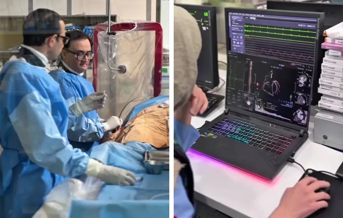
For the first time in Iran, artificial intelligence-based software has come to aid of cardiologists at Razavi Hospital to examine condition of a patient's heart vessels in greater detail prior to performing angioplasty and to map out treatment intervention plan.
Imagine a physician being able to have a precise picture of a patient's heart vessels before patient enters the angiography suite; knowing where the blockages are located, what their severity is, and what tools must be at hand to open pathway for blood supply to the heart. This is no longer merely an image of the vessels; it is a map for entering one of the most delicate medical interventions.
This event has now taken place at Razavi Hospital in Mashhad with aid of artificial intelligence technology. In a novel initiative, cardiologists of this hospital have utilized advanced software to analyze coronary computed tomography angiography (CCTA) images.
Dr. Yeganeh Mobasser, a specialist in cardiovascular diseases, emphasized a point which may seem simple at first glance, yet plays a decisive role in ultimate quality of this technology: the patient's heart rate.
She explained: "To achieve high-quality images in coronary CT angiography, patient's heart rate must drop below 65 beats per minute during imaging. For this reason, preparing the patient prior to imaging and their cooperation with the specialists of the CT angiography department is of great importance, because intelligent image analysis ultimately depends on the quality of the primary data."
Subsequently, Dr. Masoud Sajjadi, a cardiologist, considered the difference between this method and conventional CT angiography to lie in the volume of information extracted from images.
He continued: "In conventional methods, the physician is primarily faced with anatomical information of the vessels; however, the new software makes it possible to examine blockages, calcium levels, and characteristics of plaques inside the coronary vessels with greater precision."
The significance of this information becomes clearer when a physician intends to make a decision to open a blocked or narrowed vessel. Selecting appropriate tool, balloon size, stent type, and its length are among factors that matter in the success of the treatment intervention. Now, by analyzing images, the software provides a portion of this information to the specialist before the procedure begins.
In other words, prior to entering angiography suite, the physician is not merely confronted with an image of the heart vessels; rather, they can plan how to perform the intervention with a more precise understanding of the lesion's condition.
In this therapeutic experience, the work was not limited to analyzing CT angiography images. Following design of the angioplasty plan based on the software's information, the intravascular optical coherence tomography (OCT) imaging method was also utilized.
According to Dr. Sajjadi, findings obtained from OCT corresponded with the information provided by the software; a matter that demonstrates importance of this technology in providing practical information to the physician.
He cited this case as the first experience carried out in Iran using this software—an experience in which artificial intelligence technology helped design the treatment map prior to commencement of the intervention.
What matters in this initiative is not merely the addition of a new device or software to hospital equipment. The core issue is a change in the decision-making approach prior to intervention; that the physician can be better prepared to enter the patient's heart vessels with more precise information and pre-select required tools.
Given this significant development, it can be said that artificial intelligence is not going to replace the cardiologist; it is going to provide them with more details. This technology has now become part of the pathway toward more precise diagnosis and treatment of heart diseases at Razavi Hospital.
Related News


Post comments
- Editor's suggestion
- Latest news
The First Autumn Rain at the Holy Razavi Shrine
Elementary School Student of Imam Reza Schools Wins Team Gold Medal at World Inventions Exhibition
Razavi Hospital employs AI in diagnosis, treatment
Razavi Dome Draped in Black and Placement of Fatimid Inscriptions at Razavi Holy Shrine
Proposal for Establishing a Joint Iran-Uzbekistan Technology Development Office Presented
**Signing of Memorandum of Understanding on Cooperation Between Office of Deputy Custodian for International Affairs of Astan Quds Razavi and the International Foundation for Razavi Art and Culture**
The 11th International Assembly of "Combatants in Exile"
Chief Justice at the Closing Ceremony of the "Combatants in Exile" Assembly: Confronting Arrogance is an Everlasting Duty
Intl. “Mujahideen in Exile” Summit opens focusing on anti-Imperialism, legacy of Martyred Leader
Iraqi, Iranian people allies, partners in defending Islam, sacred values
Zealous tribal sheikhs, leaders of Iraq, khuzestan meet with AQR custodian
Enemy seeks to break deep bond between nations of Iran, Iraq: AQR custodian
Iraqi tribal sheikhs visit Imam Reza shrine
Iraqi tribal sheikhs renew covenant with Martyred Leader in Razavi shrine
Razavi shrine servants welcome Iraqi tribal Sheikhs at Mashhad Airport 2
AQR allocates special budget for those hit by US-Israeli crime in Lamerd
Under protection of the Imam; A visual narrative of dispatch of families of Martyrs and Wounded people of Lamerd to Mashhad
AQR honors Iraqi tribal sheikhs active in funeral procession of the Martyred Leader
Razavi shrine servants welcome Iraqi tribal Sheikhs at Mashhad Airport
AQR, Sadr International Award explore ways to expand cooperation
Turkish Shia visitors praise spiritual atmosphere and services at Imam Reza Shrine
Campaigns